

Cardiology investigations
I can arrange a comprehensive range of cardiac investigations which are tailored to the individual patient as necessary. Some of the investigations are detailed below.
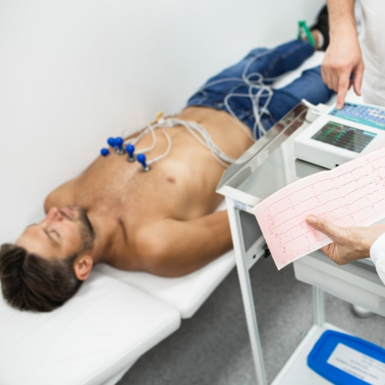
12 Lead ECG
A 12 lead ECG involves attaching a number of stickers to the chest, arms and legs. This allows a detailed electrical analysis at that point in time and can give information such as heart rate and rhythm, evidence of any heart blocks as well as pointers towards potentially structural problems with the heart.
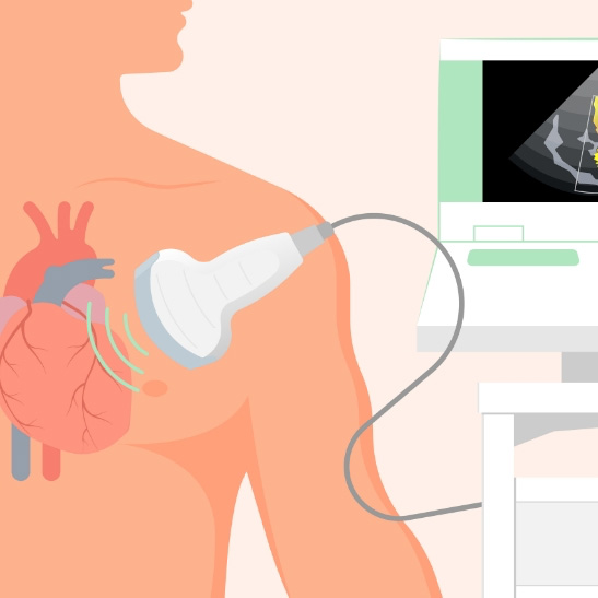
Echocardiogram
This is an ultrasound assessment of the heart and involves “looking at the heart” with an ultrasound probe and some jelly, in the same way as baby scans are performed. This gives very useful information regarding the structure and function of the heart, including sizes of the heart chambers, how the valves are working and evidence of increased pressures within the heart.
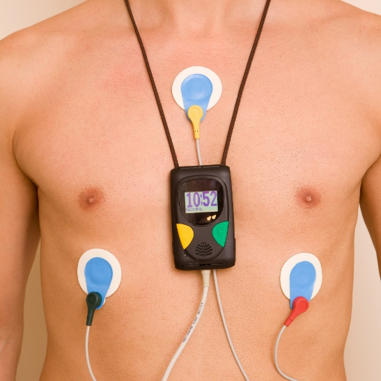
Prolonged ECG monitoring
This is useful to assess what the heart rate and rhythm is doing over a period of time – from 24 hours to 14 days. This involves wearing a monitor (more modern monitors usually involve a patch attached to the chest rather than lots of leads and stickers) as an outpatient allowing you to undertake normal activities so I can see how the heart reacts and what it is doing at the time of any symptoms.
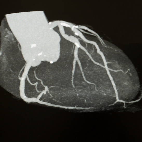
CT coronary angiogram
This uses X rays to provide information regarding the coronary arteries, and can identify calcium build up (hardening of the arteries) as well as any narrowings or blockages. This can be a very useful alternative to a conventional coronary angiogram.

Conventional coronary angiogram
This involves advancing a small tube (catheter) via one of the arteries (usually at the wrist) using local anaesthetic. X ray contrast (dye) is then injected, and X rays used to evaluate any narrowings or blockages in the arteries supplying the heart with blood.

Cardiac MRI
This type of scan uses magnets to give a very detailed picture of the heart muscle and is particularly useful in some instances such as identifying areas of scarring, or any heart muscle disorders.
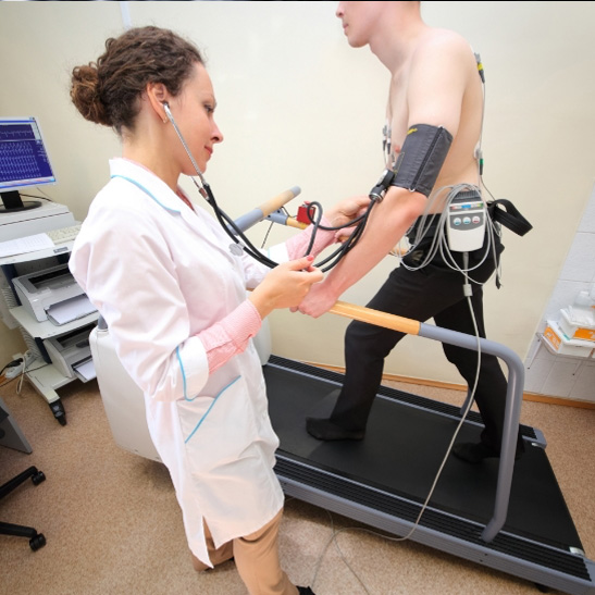
Exercise ECG
This involves evaluating the ECG when exercising, usually on a treadmill. This can give useful information as to how the heart is responding to exercise including any changes in rhythm or strain.

Exercise/Stress Echo
This involves stressing the heart using exercise (usually an exercise bike) or medication to increase the heart rate and then evaluating the heart using ultrasound to look for any changes in how the heart is working.
Transoesophageal echo
The patient swallows a thin tube which allows ultrasound pictures of the heart from the oesophagus (gullet) which is behind the heart. This has an advantage over normal echo in that the ribs are not in the way. This allows very detailed assessment of structural problems such as problems with heart valves and can help with specific treatment planning such as valve repair operations.

Sleep study
This involves monitoring the heart and breathing whilst asleep to look for any evidence of sleep apnoea which can be related to several heart problems.

“An absolute gentleman! His explanations were absolutely perfect. He listens and he answers questions without jargon or avoidance. A wonderful experience.”
— 25 Sept 2025

